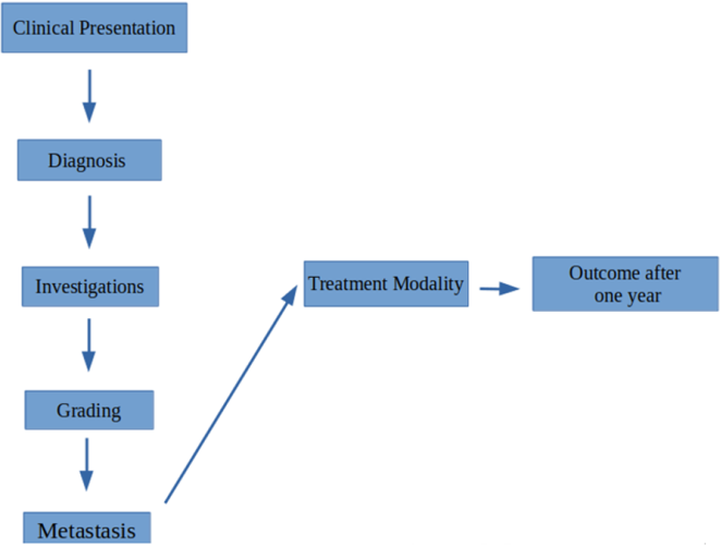
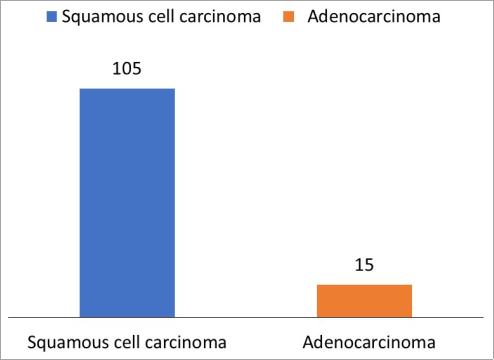
Background: Esophageal cancer is a global health concern, ranking as the seventh leading cause of cancer-related deaths. In Sudan, the rising incidence, particularly of adenocarcinoma, presents a significant challenge. Limited treatment resources affect patient outcomes, despite surgical management for localized disease and systemic therapy for metastatic cases. Early detection is crucial for improving prognosis. This study investigates the presentation patterns and management strategies for esophageal cancer within a Sudanese context. Methodology: A retrospective, cross-sectional study was conducted at the Tumor Therapy and Cancer Research Center in Sudan, analyzing 120 esophageal cancer patient records from 2016 to 2023. Data encompassed demographics, clinical presentations, diagnostic findings, treatment modalities, and patient outcomes. Statistical analysis was performed using SPSS. Ethical approvals were obtained prior to data collection. Results: Among the 120 patients, 105 had squamous cell carcinoma (SCC) and 15 had adenocarcinoma (AC). The majority of patients were between 51–60 years (25.8%) and over 70 years (22.5%). More than half (61.7%) were female, and most resided in rural areas (77.5%). Helicobacter pylori infection (17.5%) was the most common risk factor, followed by smoking (10.8%). Other reported risk factors included gastroesophageal reflux disease, alcohol use, and a family history of esophageal cancer. Dysphagia was the most common presenting symptom, with most patients diagnosed at an advanced stage. Conclusion: SCC predominantly affected older females, while AC was more common in younger males. Dysphagia was the main presenting symptom. Low utilization of neoadjuvant therapy and higher mortality rates in AC patients (60% vs. 30.5% in SCC) highlight the need for improved screening, early diagnosis, optimized treatment protocols.
| Published in | Pathology and Laboratory Medicine (Volume 9, Issue 2) |
| DOI | 10.11648/j.plm.20250902.11 |
| Page(s) | 38-48 |
| Creative Commons |
This is an Open Access article, distributed under the terms of the Creative Commons Attribution 4.0 International License (http://creativecommons.org/licenses/by/4.0/), which permits unrestricted use, distribution and reproduction in any medium or format, provided the original work is properly cited. |
| Copyright |
Copyright © The Author(s), 2025. Published by Science Publishing Group |
Esophageal Cancer, Sudan, Adenocarcinoma, Squamous Cell Carcinoma, Dysphagia, Mortality, Neoadjuvant Therapy, Retrospective Study
Characteristics | Frequency | Percent |
|---|---|---|
Age: | ||
20-30 years | 1 | 0.8 |
31-40 years | 17 | 14.2 |
41-50 years | 18 | 15.0 |
51-60 years | 31 | 25.8 |
61-70 years | 26 | 21.7 |
more than 70 years | 27 | 22.5 |
Sex: | ||
Male | 46 | 38.3 |
Female | 74 | 61.7 |
Residence: | ||
Urban | 27 | 22.5 |
Rural | 93 | 77.5 |
Occupation: | ||
Housewife | 71 | 59.2 |
Employee | 8 | 6.7 |
Free worker | 38 | 31.7 |
Unemployed | 3 | 2.5 |
Marital: | ||
Single | 14 | 11.7 |
Married | 79 | 65.8 |
Divorced | 6 | 5.0 |
Widow | 21 | 17.5 |
Risk factors: | ||
Gastroesophageal reflex | 2 | 1.7 |
History of H. Pylori infection | 21 | 17.5 |
Frequent use of alcohol | 3 | 2.5 |
Family history | 4 | 3.3 |
Smoking | 13 | 10.8 |
Patients’ characteristics | Squamous cell carcinoma | Adenocarcinoma |
|---|---|---|
Age groups: | ||
20-30 years | 1.0 (1.0%) | 0.0 (0.0%) |
31-40 years | 16.0 (15.2%) | 1.0 (6.7%) |
41-50 years | 18.0 (17.1%) | 0.0 (0.0%) |
51-60 years | 24.0 (22.9%) | 7.0 (46.7%) |
61-70 years | 19.0 (18.1%) | 7.0 (46.7%) |
More than 70 years | 27.0 (25.7%) | 0.0 (0.0%) |
Sex: | ||
Male | 35.0 (33.3%) | 11.0 (73.3%) |
Female | 70.0 (66.7%) | 4.0 (26.7%) |
Residence: | ||
Urban | 24.0 (22.9%) | 3.0 (20.0%) |
Rural | 81.0 (77.1%) | 12.0 (80.0%) |
Occupation: | ||
Housewife | 68.0 (64.8%) | 3.0 (20.0%) |
Employee | 6.0 (5.7%) | 2.0 (13.3%) |
Free worker | 28.0 (26.7%) | 10.0 (66.7%) |
Unemployed | 3.0 (2.9%) | 0.0 (0.0%) |
Marital status: | ||
Single | 14.0 (13.3%) | 0.0 (0.0%) |
Married | 65.0 (61.9%) | 14.0 (93.3%) |
Divorced | 5.0 (4.8%) | 1.0 (6.7%) |
Widow | 21.0 (20.0%) | 0.0 (0.0%) |
Risk factors: | ||
GEARD | 2.0 (1.9%) | 0.0 (0.0%) |
History of Pylori infection | 14.0 (13.3%) | 7.0 (46.7%) |
Frequent use of alcohol | 2.0 (1.9%) | 1.0 (6.7%) |
Family history | 3.0 (2.9%) | 1.0 (6.7%) |
Smoking | 11.0 (10.5%) | 2.0 (13.3%) |
Tumor characteristics | Squamous cell carcinoma | Adenocarcinoma |
|---|---|---|
Clinical presentation: | ||
Dysphagia | 97.0 (92.4%) | 13.0 (86.7%) |
Hoarseness of voice | 3.0 (2.9%) | 0.0 (0.0%) |
Odynophagia | 9.0 (8.6%) | 0.0 (0.0%) |
Vomiting | 12.0 (11.4%) | 5.0 (33.3%) |
Hematemesis | 4.0 (3.8%) | 0.0 (0.0%) |
Weight loss | 34.0 (32.4%) | 5.0 (33.3%) |
Neck swelling | 1.0 (1.0%) | 0.0 (0.0%) |
Recurrent chest infection | 8.0 (7.6%) | 0.0 (0.0%) |
Excessive salivation | 2.0 (1.9%) | 0.0 (0.0%) |
Epigastric pain | 10.0 (9.5%) | 5.0 (33.3%) |
Site of mass: | ||
Upper third | 23.0 (21.9%) | 0.0 (0.0%) |
Middle third | 39.0 (37.1%) | 1.0 (6.7%) |
Lower third | 43.0 (41.0%) | 14.0 (93.3%) |
Grade at presentation: | ||
Stage I | 38.0 (36.2%) | 7.0 (46.7%) |
Stage II | 46.0 (43.8%) | 5.0 (33.3%) |
Stage III | 17.0 (16.2%) | 2.0 (13.3%) |
Stage IV | 4.0 (3.8%) | 1.0 (6.7%) |
Metastasis: | ||
yes | 27.0 (25.7%) | 8.0 (53.3%) |
no | 78.0 (74.3%) | 7.0 (46.7%) |
Site of metastasis: | ||
Lung | 17.0 (45.9%) | 4.0 (50.0%) |
Bone | 2.0 (5.4%) | 0.0 (0.0%) |
Liver | 16.0 (43.2%) | 3.0 (37.5%) |
Brain | 0.0 (0.0%) | 1.0 (12.5%) |
Others | 2.0 (5.4%) | 0.0 (0.0%) |
O. G. D findings: | ||
Mass | 60.0 (57.1%) | 8.0 (53.3%) |
Fungating mass | 32.0 (30.5%) | 4.0 (26.7%) |
Ulcerative mass | 7.0 (6.7%) | 1.0 (6.7%) |
Stricture | 6.0 (5.7%) | 2.0 (13.3%) |
Variables | Squamous cell carcinoma | Adenocarcinoma |
|---|---|---|
Neo-Adjuvant Therapy: | ||
Received neo-adjuvant therapy | 3.0 (2.9%) | 1.0 (6.7%) |
Did not receive neo-adjuvant therapy | 102.0 (97.1%) | 14.0 (93.3%) |
Surgical Intervention: | ||
No surgery performed | 105.0 (100.0%) | 15.0 (100.0%) |
Nutritional Support: | ||
Percutaneous endoscopic gastrostomy (PEG) tube | 1.0 (1.0%) | 0.0 (0.0%) |
Open gastrostomy | 2.0 (1.9%) | 0.0 (0.0%) |
Open jejunostomy | 6.0 (5.7%) | 0.0 (0.0%) |
Nasogastric (NG) tube | 4.0 (3.8%) | 1.0 (6.7%) |
No nutritional support provided | 92.0 (87.6%) | 14.0 (93.3%) |
Adjuvant Therapy: | ||
Received adjuvant therapy | 96.0 (91.4%) | 14.0 (93.3%) |
Did not receive adjuvant therapy | 9.0 (8.6%) | 1.0 (6.7%) |
One-Year Outcome | ||
Cured | 1.0 (1.0%) | 1.0 (6.7%) |
Under treatment | 62.0 (59.0%) | 4.0 (26.7%) |
Developed metastasis | 10.0 (9.5%) | 1.0 (6.7%) |
Death | 32.0 (30.5%) | 9.0 (60.0%) |
Variable | Metastasis Present (n=35) | Metastasis Absent (n=85) | p-value |
|---|---|---|---|
Age Group (Years) | |||
20-30 | 0.0 (0.0%) | 1.0 (1.2%) | |
31-40 | 6.0 (17.1%) | 11.0 (12.9%) | |
41-50 | 5.0 (14.3%) | 13.0 (15.3%) | |
51-60 | 12.0 (34.3%) | 19.0 (22.4%) | 0.095 |
61-70 | 10.0 (28.6%) | 16.0 (18.8%) | |
> 70 | 2.0 (5.7%) | 25.0 (29.4%) | |
Sex | |||
Male | 19.0 (54.3%) | 27.0 (31.8%) | |
Female | 16.0 (45.7%) | 58.0 (68.2%) | 0.021 |
Occupation | |||
Housewife | 14.0 (40.0%) | 57.0 (67.1%) | |
Employee | 1.0 (2.9%) | 7.0 (8.2%) | |
Free worker | 19.0 (54.3%) | 19.0 (22.4%) | 0.007 |
Unemployed | 1.0 (2.9%) | 2.0 (2.4%) | |
Histological Type | |||
Squamous cell carcinoma | 27.0 (77.1%) | 78.0 (91.8%) | |
Adenocarcinoma | 8.0 (22.9%) | 7.0 (8.2%) | 0.028 |
Tumor Stage at Presentation | |||
Stage I | 9.0 (25.7%) | 36.0 (42.4%) | |
Stage II | 13.0 (37.1%) | 38.0 (44.7%) | |
Stage III | 9.0 (25.7%) | 10.0 (11.8%) | 0.009 |
Stage IV | 4.0 (11.4%) | 1.0 (1.2%) | |
Tumor Location | |||
Upper third | 5.0 (14.3%) | 18.0 (21.2%) | |
Middle third | 7.0 (20.0%) | 33.0 (38.8%) | 0.035 |
Lower third | 23.0 (65.7%) | 34.0 (40.0%) | |
Neo-Adjuvant Therapy | |||
Received | 2.0 (5.7%) | 2.0 (2.4%) | |
Not received | 33.0 (94.3%) | 83.0 (97.6%) | 0.351 |
Adjuvant Therapy | |||
Received | 34.0 (97.1%) | 76.0 (89.4%) | |
Not received | 1.0 (2.9%) | 9.0 (10.6%) | 0.164 |
Predictor | B | S. E. | Wald | df | Sig. | Exp (B) | 95% confidence level | |
|---|---|---|---|---|---|---|---|---|
lower | upper | |||||||
Age | 0.201 | 0.178 | 1.286 | 1 | 0.257 | 1.223 | 0.864 | 1.732 |
Sex | -0.277 | 1.047 | .070 | 1 | 0.792 | 0.758 | 0.097 | 5.908 |
Occupation | -0.624 | 0.522 | 1.428 | 1 | 0.232 | 0.536 | 0.193 | 1.491 |
Marital status | 0.406 | 0.319 | 1.619 | 1 | 0.203 | 1.501 | 0.803 | 2.807 |
Histological Type | 0.836 | 0.654 | 1.633 | 1 | 0.201 | 2.308 | 0.640 | 8.324 |
Tumor Grade at Presentation | -0.789 | 0.289 | 7.469 | 1 | 0.006 | 0.454 | 0.258 | 0.800 |
Tumor Location | -0.309 | 0.319 | 0.935 | 1 | 0.334 | 0.735 | 0.393 | 1.373 |
Constant | 1.501 | 3.066 | 0.240 | 1 | 0.624 | 4.486 | ||
AC | Adenocarcinoma |
ADC | Adenocarcinoma |
EC | Esophageal Cancer |
ESCC | Esophageal Squamous-cell Carcinoma |
GERD | Gastroesophageal Reflux Disease |
GRCC | Regional Cancer Center |
HBH | High-burden Hospitals |
ODG | Esophagogastroduodenoscopy |
RFS | Recurrence-Free Survival |
SSC | Squamous Cell Carcinoma |
| [1] | Sung H, Ferlay J, Siegel RL, Laversanne M, Soerjomataram I, Jemal A, et al. Global Cancer Statistics 2020: GLOBOCAN Estimates of Incidence and Mortality Worldwide for 36 Cancers in 185 Countries. CA Cancer J Clin. 2021; 71(3): 209–49. |
| [2] | The global, regional, and national burden of oesophageal cancer and its attributable risk factors in 195 countries and territories, 1990–2017: a systematic analysis for the Global Burden of Disease Study 2017. Lancet Gastroenterol Hepatol. 2020 Apr 1; 5(6): 582–97. |
| [3] | Stawinski PM, Dziadkowiec KN, Kuo LA, Echavarria J, Saligram S. Barrett’s Esophagus: An Updated Review. Diagnostics. 2023 Jan; 13(2): 321. |
| [4] | Saeed MEM, Cao J, Fadul B, Kadioglu O, Khalid HE, Yassin Z, et al. A Five-year Survey of Cancer Prevalence in Sudan. Anticancer Res. 2016 Jan 1; 36(1): 279–86. |
| [5] | Musa IH, Musa TH, Musa HH, Ahmed ME. Esophageal cancer epidemiology, diagnosis, and management in Sudan - A review. Med J Malaysia. 2021 Sep; 76(5): 691–7. |
| [6] | Saeed IE, Weng HY, Mohamed KH, Mohammed SI. Cancer incidence in Khartoum, Sudan: first results from the Cancer Registry, 2009-2010. Cancer Med. 2014 Aug; 3(4): 1075–84. |
| [7] | Ahmed ME, Mahadi SI, Ali BM. The surgical treatment of esophageal cancer in Sudan: A 100 consecutive cases. Int J Surg Lond Engl. 2016 May; 29: 101–7. |
| [8] | Deboever N, Jones CM, Yamashita K, Ajani JA, Hofstetter WL. Advances in diagnosis and management of cancer of the esophagus. BMJ. 2024 Jun 3; 385: e074962. |
| [9] | Markar S, Gronnier C, Duhamel A, Bigourdan JM, Badic B, du Rieu MC, et al. Pattern of Postoperative Mortality After Esophageal Cancer Resection According to Center Volume: Results from a Large European Multicenter Study. Ann Surg Oncol. 2015 Aug 1; 22(8): 2615–23. |
| [10] | Rafiemanesh H, Maleki F, Mohammadian-Hafshejani A, Salemi M, Salehiniya H. The Trend in Histological Changes and the Incidence of Esophagus Cancer in Iran (2003–2008). Int J Prev Med. 2016 Feb 8; 7: 31. |
| [11] | Wheeler JB, Reed CE. Epidemiology of esophageal cancer. Surg Clin North Am. 2012 Oct; 92(5): 1077–87. |
| [12] | Bollschweiler E, Wolfgarten E, Nowroth T, Rosendahl U, Mönig SP, Hölscher AH. Vitamin intake and risk of subtypes of esophageal cancer in Germany. J Cancer Res Clin Oncol. 2002 Oct; 128(10): 575–80. |
| [13] | Lim RZM, Mahendran HA, Malaysian Upper Gastrointestinal Surgical Society (MUGIS). Esophageal squamous cell carcinoma and adenocarcinoma in Malaysia - Pooled data from upper gastrointestinal centers in a multiethnic Asian population. Cancer Epidemiol. 2022 Oct; 80: 102211. |
| [14] | Chong VH, Telisinghe PU, Chong CF. Esophageal Cancer in Brunei Darussalam over a three Decade Period: an Epidemiologic Study of Trends and Differences between Genders and Racial Groups. Asian Pac J Cancer Prev APJCP. 2015; 16(9): 4123–6. |
| [15] | Shin A, Won YJ, Jung HK, Kong HJ, Jung KW, Oh CM, et al. Trends in incidence and survival of esophageal cancer in Korea: Analysis of the Korea Central Cancer Registry Database. J Gastroenterol Hepatol. 2018 Dec; 33(12): 1961–8. |
| [16] | Koizumi S, Motoyama S, Iijima K. Is the incidence of esophageal adenocarcinoma increasing in Japan? Trends from the data of a hospital-based registration system in Akita Prefecture, Japan. J Gastroenterol. 2018 Jul; 53(7): 827–33. |
| [17] | Yusupbekov A, Kanda M, Usmanov B, Tuychiev O, Baymakov S, Sakamoto J, et al. Surveillance of Esophageal Cancer in the Republic of Uzbekistan from 2000 to 2018. Asian Pac J Cancer Prev APJCP. 2020 Aug 1; 21(8): 2281–5. |
| [18] | Noronha V, Sekar A, Rajendra A, Mokal S, Patil V, Menon N, et al. Epidemiological Trend of Esophageal Cancer at a Tertiary Cancer Center in Mumbai, India, Over the Past 15 Years. J Gastrointest Cancer. 2023 Sep; 54(3): 903–12. |
| [19] | Eroğlu A, Aydın Y, Altuntaş B, Gündoğdu B, Yılmaz Ö. The increasing incidence of esophageal squamous cell carcinoma in women in Turkey. Turk J Med Sci. 2016 Nov 17; 46(5): 1443–8. |
| [20] | Asombang AW, Kasongo N, Muyutu J, Montiero JFG, Chishinga N, Chipaila J, et al. Descriptive analysis of esophageal cancer in Zambia using the cancer disease hospital database: young age, late stage at presentation. Pan Afr Med J. 2021; 39: 12. |
| [21] | Abdihamid O, Abdourahman H, Ibrahim A, Kareu T, Hadi A, Omar A, et al. Landscape of esophageal cancer in Northern Kenya: experience from Garissa Regional Cancer Center. Ecancermedicalscience. 2024; 18: 1694. |
| [22] | González Ortiz DI, Toro DH. Esophageal cancer subtypes and survival rates at the VA Caribbean Healthcare System: a 10-year experience. Boletin Asoc Medica P R. 2009; 101(3): 14–7. |
| [23] | Fagundes RB, de Carli D, Xaubet RV, Cantarelli JC. Unchanging pattern of prevalence of esophageal cancer, overall and by histological subtype, in the endoscopy service of the main referral hospital in the central region of Rio Grande do Sul State, in Southern Brazil. Dis Esophagus Off J Int Soc Dis Esophagus. 2016 Aug; 29(6): 603–6. |
| [24] | He H, Chen N, Hou Y, Wang Z, Zhang Y, Zhang G, et al. Trends in the incidence and survival of patients with esophageal cancer: A SEER database analysis. Thorac Cancer. 2020 May; 11(5): 1121–8. |
| [25] | Njei B, McCarty TR, Birk JW. Trends in esophageal cancer survival in United States adults from 1973 to 2009: A SEER database analysis. J Gastroenterol Hepatol. 2016 Jun; 31(6): 1141–6. |
| [26] | Henry MAC de A, Lerco MM, Ribeiro PW, Rodrigues MAM. Epidemiological features of esophageal cancer. Squamous cell carcinoma versus adenocarcinoma. Acta Cir Bras. 2014 Jun; 29(6): 389–93. |
| [27] | Matz M, Valkov M, Šekerija M, Luttman S, Caldarella A, Coleman MP, et al. Worldwide trends in esophageal cancer survival, by sub-site, morphology, and sex: an analysis of 696,974 adults diagnosed in 60 countries during 2000-2014 (CONCORD-3). Cancer Commun Lond Engl. 2023 Sep; 43(9): 963–80. |
| [28] | Kauppila JH, Mattsson F, Brusselaers N, Lagergren J. Prognosis of oesophageal adenocarcinoma and squamous cell carcinoma following surgery and no surgery in a nationwide Swedish cohort study. BMJ Open. 2018 May 10; 8(5): e021495. |
| [29] | Sridhar P, Bhatt M, Qureshi MM, Asokan S, Truong MT, Suzuki K, et al. Esophageal Cancer Presentation, Treatment, and Outcomes Vary With Hospital Safety-Net Burden. Ann Thorac Surg. 2019 May; 107(5): 1472–9. |
| [30] | Du KN, Feng L, Newhouse A, Mehta J, Lasala J, Mena GE, et al. Effects of Intraoperative Opioid Use on Recurrence-Free and Overall Survival in Patients With Esophageal Adenocarcinoma and Squamous Cell Carcinoma. Anesth Analg. 2018 Jul; 127(1): 210–6. |
APA Style
Awad, A. M., Abdelgadir, H. S., Mostafa, M. M., Elmagboul, M. E., Ahmed, A. K., et al. (2025). Pattern of Presentations and Management of Esophageal Cancer in Tumor Therapy and Cancer Research Center 2024. Pathology and Laboratory Medicine, 9(2), 38-48. https://doi.org/10.11648/j.plm.20250902.11
ACS Style
Awad, A. M.; Abdelgadir, H. S.; Mostafa, M. M.; Elmagboul, M. E.; Ahmed, A. K., et al. Pattern of Presentations and Management of Esophageal Cancer in Tumor Therapy and Cancer Research Center 2024. Pathol. Lab. Med. 2025, 9(2), 38-48. doi: 10.11648/j.plm.20250902.11
AMA Style
Awad AM, Abdelgadir HS, Mostafa MM, Elmagboul ME, Ahmed AK, et al. Pattern of Presentations and Management of Esophageal Cancer in Tumor Therapy and Cancer Research Center 2024. Pathol Lab Med. 2025;9(2):38-48. doi: 10.11648/j.plm.20250902.11
@article{10.11648/j.plm.20250902.11,
  author = {Ahmed Mahjoub Awad and Hiba Salah Abdelgadir and Maram Mohamed Mostafa and Maram Elnair Elmagboul and Afra Kamal Ahmed and Mohammed Hammad Jaber and Aymen Khalid Mudawe and Hind Salah Abdelgadir and Lea Nohra and Fatima Abdallh and Mosab Abdelgader Ahmed},
  title = {Pattern of Presentations and Management of Esophageal Cancer in Tumor Therapy and Cancer Research Center 2024},
  journal = {Pathology and Laboratory Medicine},
  volume = {9},
  number = {2},
  pages = {38-48},
  doi = {10.11648/j.plm.20250902.11},
  url = {https://doi.org/10.11648/j.plm.20250902.11},
  eprint = {https://article.sciencepublishinggroup.com/pdf/10.11648.j.plm.20250902.11},
  abstract = {Background: Esophageal cancer is a global health concern, ranking as the seventh leading cause of cancer-related deaths. In Sudan, the rising incidence, particularly of adenocarcinoma, presents a significant challenge. Limited treatment resources affect patient outcomes, despite surgical management for localized disease and systemic therapy for metastatic cases. Early detection is crucial for improving prognosis. This study investigates the presentation patterns and management strategies for esophageal cancer within a Sudanese context. Methodology: A retrospective, cross-sectional study was conducted at the Tumor Therapy and Cancer Research Center in Sudan, analyzing 120 esophageal cancer patient records from 2016 to 2023. Data encompassed demographics, clinical presentations, diagnostic findings, treatment modalities, and patient outcomes. Statistical analysis was performed using SPSS. Ethical approvals were obtained prior to data collection. Results: Among the 120 patients, 105 had squamous cell carcinoma (SCC) and 15 had adenocarcinoma (AC). The majority of patients were between 51–60 years (25.8%) and over 70 years (22.5%). More than half (61.7%) were female, and most resided in rural areas (77.5%). Helicobacter pylori infection (17.5%) was the most common risk factor, followed by smoking (10.8%). Other reported risk factors included gastroesophageal reflux disease, alcohol use, and a family history of esophageal cancer. Dysphagia was the most common presenting symptom, with most patients diagnosed at an advanced stage. Conclusion: SCC predominantly affected older females, while AC was more common in younger males. Dysphagia was the main presenting symptom. Low utilization of neoadjuvant therapy and higher mortality rates in AC patients (60% vs. 30.5% in SCC) highlight the need for improved screening, early diagnosis, optimized treatment protocols.},
 year = {2025}
}
TY  - JOUR
T1  - Pattern of Presentations and Management of Esophageal Cancer in Tumor Therapy and Cancer Research Center 2024
AU  - Ahmed Mahjoub Awad
AU  - Hiba Salah Abdelgadir
AU  - Maram Mohamed Mostafa
AU  - Maram Elnair Elmagboul
AU  - Afra Kamal Ahmed
AU  - Mohammed Hammad Jaber
AU  - Aymen Khalid Mudawe
AU  - Hind Salah Abdelgadir
AU  - Lea Nohra
AU  - Fatima Abdallh
AU  - Mosab Abdelgader Ahmed
Y1  - 2025/10/09
PY  - 2025
N1  - https://doi.org/10.11648/j.plm.20250902.11
DO  - 10.11648/j.plm.20250902.11
T2  - Pathology and Laboratory Medicine
JF  - Pathology and Laboratory Medicine
JO  - Pathology and Laboratory Medicine
SP  - 38
EP  - 48
PB  - Science Publishing Group
SN  - 2640-4478
UR  - https://doi.org/10.11648/j.plm.20250902.11
AB  - Background: Esophageal cancer is a global health concern, ranking as the seventh leading cause of cancer-related deaths. In Sudan, the rising incidence, particularly of adenocarcinoma, presents a significant challenge. Limited treatment resources affect patient outcomes, despite surgical management for localized disease and systemic therapy for metastatic cases. Early detection is crucial for improving prognosis. This study investigates the presentation patterns and management strategies for esophageal cancer within a Sudanese context. Methodology: A retrospective, cross-sectional study was conducted at the Tumor Therapy and Cancer Research Center in Sudan, analyzing 120 esophageal cancer patient records from 2016 to 2023. Data encompassed demographics, clinical presentations, diagnostic findings, treatment modalities, and patient outcomes. Statistical analysis was performed using SPSS. Ethical approvals were obtained prior to data collection. Results: Among the 120 patients, 105 had squamous cell carcinoma (SCC) and 15 had adenocarcinoma (AC). The majority of patients were between 51–60 years (25.8%) and over 70 years (22.5%). More than half (61.7%) were female, and most resided in rural areas (77.5%). Helicobacter pylori infection (17.5%) was the most common risk factor, followed by smoking (10.8%). Other reported risk factors included gastroesophageal reflux disease, alcohol use, and a family history of esophageal cancer. Dysphagia was the most common presenting symptom, with most patients diagnosed at an advanced stage. Conclusion: SCC predominantly affected older females, while AC was more common in younger males. Dysphagia was the main presenting symptom. Low utilization of neoadjuvant therapy and higher mortality rates in AC patients (60% vs. 30.5% in SCC) highlight the need for improved screening, early diagnosis, optimized treatment protocols.
VL  - 9
IS  - 2
ER  -
Surgery Department, Shendi University, Shendi, Sudan
Family Medicine, Community Medicine Department, Alzaiem Alazhari University, Khartoum, Sudan
Surgery Department, Shendi University, Shendi, Sudan
Surgery Department, Shendi University, Shendi, Sudan
Faculty of Medicine, Alzaiem Alazhari University, Khartoum, Sudan
Faculty of Medicine, Alzaiem Alazhari University, Khartoum, Sudan
Faculty of Medicine, Alzaiem Alazhari University, Khartoum, Sudan
Faculty of Medicine, Alzaiem Alazhari University, Khartoum, Sudan
Faculty of Medical Sciences, Lebanese University, Beirut, Lebanon
Faculty of Medicine, Hashemite University, Zarqa, Jordan
Faculty of Clinical Pharmacy, Khartoum College of Medical Sciences, Khartoum, Sudan